 |
Organic Compound Identification Using Infrared Spectroscopy
Dr. Walt Volland, Bellevue Community College
All rights reserved 1999, Bellevue, Washington
Description
This exercise is intended to familiarize you with the identification of functional groups in organic compounds using infrared spectra. Before you can use this technique, you need to have an introduction to infrared spectroscopy and to what an IR spectrum is.
Infrared spectroscopy deals with the interaction of infrared light with matter. The energy of an infrared photon can be calculated using the Planck energy relation.
where h = 6.6 x
10-34
joule second and
n = frequency
of the photon. This shows that high energy photons have high
frequency.
The frequency, n, and speed of light, c, are related through the relation
where c = 3.0 x 10⁸
meter/second and l
= wavelength for the light

These two equations can be used to identify
a common spectroscopic unit called wavenumber, ![]() ,
which is the reciprocal of the wavelength.
,
which is the reciprocal of the wavelength.
![]() = wavenumber =
= wavenumber = ![]() has units of (cm-1)
has units of (cm-1)
You can see that both frequency and wavenumber are directly proportional to energy.
Molecules are flexible, moving collections of atoms. The atoms in a molecule are constantly oscillating around average positions. Bond lengths and bond angles are continuously changing due to this vibration. A molecule absorbs infrared radiation when the vibration of the atoms in the molecule produces an oscillating electric field with the same frequency as the frequency of incident IR "light".
All of the motions can be described in terms
of two types of molecular vibrations. One type of vibration, a
stretch, produces a change of bond length. A stretch is a rhythmic
movement along the line between the atoms so that the interatomic
distance is either increasing or decreasing. ![]()
The second type of vibration, a bend, results in a change in bond angle. These are also sometimes called scissoring, rocking, or "wig wag" motions.

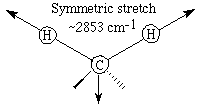
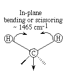
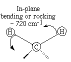
Each of these two main types of vibration can have variations. A stretch can be symmetric or asymmetric. Bending can occur in the plane of the molecule or out of plane; it can be scissoring, like blades of a pair of scissors, or rocking, where two atoms move in the same direction.
Different stretching and bending vibrations can be visualized by considering the CH2 group in hydrocarbons. The arrows indicate the direction of motion. The stretching motions require more energy than the bending ones.


Note the high wavenumber (high energy) required to produce these motions.
The bending motions are sometimes described as wagging or scissoring motions.


You can see that the lower wavenumber values are consistent with lower energy to cause these vibrations.
A molecule absorbs a unique set of IR light frequencies. Its IR spectrum is often likened to a person's fingerprints. These frequencies match the natural vibrational modes of the molecule. A molecule absorbs only those frequencies of IR light that match vibrations that cause a change in the dipole moment of the molecule. Bonds in symmetric N2 and H2 molecules do not absorb IR because stretching does not change the dipole moment, and bending cannot occur with only 2 atoms in the molecule. Any individual bond in an organic molecule with symmetric structures and identical groups at each end of the bond will not absorb in the IR range. For example, in ethane, the bond between the carbon atoms does not absorb IR because there is a methyl group at each end of the bond. The C-H bonds within the methyl groups do absorb.
In a complicated molecule many fundamental vibrations are possible, but not all are observed. Some motions do not change the dipole moment for the molecule; some are so much alike that they coalesce into one band.
Even though an IR spectrum is characteristic
for an entire molecule, there are certain groups of atoms in a
molecule that give rise to absorption bands at or near the same
wavenumber, ![]() ,
,
(frequency) regardless of the rest of the structure of the molecule. These persistent characteristic bands enable you to identify major structural features of the molecule after a quick inspection of the spectrum and the use of a correlation table. The correlation table is a listing of functional groups and their characteristic absorption frequencies.
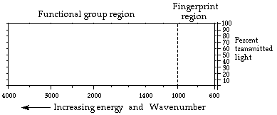
The infrared spectrum for a molecule is a graphical display. It shows the frequencies of IR radiation absorbed and the % of the incident light that passes through the molecule without being absorbed. The spectrum has two regions. The fingerprint region is unique for a molecule and the functional group region is similar for molecules with the same functional groups.

The nonlinear horizontal axis has units of wavenumbers. Each wavenumber value matches a particular frequency of infrared light. The vertical axis shows % transmitted light. At each frequency the % transmitted light is 100% for light that passes through the molecule with no interactions; it has a low value when the IR radiation interacts and excites the vibrations in the molecule.
A portion of the spectrum where % transmittance drops to a low value then rises back to near 100% is called a "band". A band is associated with a particular vibration within the molecule. The width of a band is described as broad or narrow based on how large a range of frequencies it covers. The efficiencies for the different vibrations determine how "intense" or strong the absorption bands are. A band is described as strong, medium, or weak depending on its depth.
In the hexane spectrum below the band for
the CH stretch is strong and that for the CH bend is medium. The
alkane, hexane (C6H14)
gives an IR spectrum that has relatively few bands because there are
only CH bonds that can stretch or bend. There are bands for CH
stretches at about 3000 cm-1. The CH2 bend band
appears at approximately 1450 cm-1 and the
CH3
bend at about 1400
cm-1. The spectrum also shows that shapes of bands can
differ. 
Procedure
Every molecule will have its own characteristic spectrum. The bands that appear depend on the types of bonds and the structure of the molecule. Study the sample spectra below, noting similarities and differences, and relate these to structure and bonding within the molecules.
The spectrum for the alkene, 1-hexene, C6H12, has few strong absorption bands. The spectrum has the various CH stretch bands that all hydrocarbons show near 3000 cm-1. There is a weak alkene CH stretch above 3000 cm-1. This comes from the C&emdash;H bonds on carbons 1 and 2, the two carbons that are held together by the double bond. The strong CH stretch bands below 3000 cm-1 come from carbon-hydrogen bonds in the CH2 and CH3 groups. There is an out-of-plane CH bend for the alkene in the range 1000-650 cm-1. There is also an alkene CC double bond stretch at about 1650 cm-1 .

The spectrum for cyclohexene, (C6H10) also has few strong bands. The main band is a strong CH stretch from the CH2 groups at about 3000 cm-1. The CH stretch for the alkene CH is, as always, to the left of 3000 cm-1. The CH2 bend appears at about 1450 cm-1. The other weaker bands in the range 1000-650 cm-1 are for the out of plane CH bending . There is a very weak alkene CC double bond stretch at about 1650 cm-1.

The IR spectrum for benzene, C6H6, has only four prominent bands because it is a very symmetric molecule. Every carbon has a single bond to a hydrogen. Each carbon is bonded to two other carbons and the carbon-carbon bonds are alike for all six carbons. The molecule is planar. The aromatic CH stretch appears at 3100-3000 cm-1 There are aromatic CC stretch bands (for the carbon-carbon bonds in the aromatic ring) at about 1500 cm-1. Two bands are caused by bending motions involving carbon-hydrogen bonds. The bands for CH bends appear at approximately 1000 cm-1 for the in-plane bends and at about 675 cm-1 for the out-of-plane bend.

The IR spectrum for the alcohol, ethanol
(CH3CH2OH),
is more complicated. It has a CH stretch, an OH stretch, a CO stretch
and various bending vibrations. The important point to learn here is
that no matter what alcohol molecule you deal with, the OH stretch
will appear as a broad band at approximately 3300-3500
cm-1. Likewise the CH stretch still appears at about 3000
cm-1.
The spectrum for the aldehyde, octanal (CH3(CH2)6CHO), is shown here. The most important features of the spectrum are carbonyl CO stretch near 1700 cm-1 and the CH stretch at about 3000 cm-1. If you see an IR spectrum with an intense strong band near 1700 cm-1 and the compound contains oxygen, the molecule most likely contains a carbonyl group,
![]()

The spectrum for the ketone, 2-pentanone, appears below. It also has a characteristic carbonyl band at 1700 cm-1. The CH stretch still appears at about 3000 cm-1, and the CH2 bend shows up at approximately 1400 cm-1. You can see the strong carbonyl CO stretch at approximately 1700 cm-1. You can also see that this spectrum is different from the spectrum for octanal. At this point in your study of IR spectroscopy, you can't tell which compound is an aldehyde and which is a ketone. You can tell that both octanal and a 2-pentanone contain C-H bonds and a carbonyl group.


Carboxylic acids have spectra that are even more involved. They typically have three bands caused by bonds in the COOH functional group. The band near 1700 cm-1 is due to the CO double bond. The broad band centered in the range 2700-3300 cm-1 is caused by the presence of the OH and a band near 1400 cm-1 comes from the CO single bond . The spectrum for the carboxylic acid, diphenylacetic acid, appears below. Although the aromatic CH bands complicate the spectrum, you can still see the broad OH stretch between 2700-3300 cm-1. It overlaps the CH stretch which appears near 3000 cm-1. A strong carbonyl CO stretch band exists near 1700 cm-1. The CO single bond stretch shows up near 1200 cm-1.

The spectrum for 1-bromobutane, C4H9Br, is shown here. This is relatively simple because there are only CH single bonds and the CBr bond. The CH stretch still appears at about 3000 cm-1. The CH2 bend shows up near 1400 cm-1, and you can see the CBr stretch band at approximately 700 cm-1.

IR spectra can be used to identify molecules by recording the spectrum for an unknown and comparing this to a library or data base of spectra of known compounds. Computerized spectra data bases and digitized spectra are used routinely in this way in research, medicine, criminology, and a number of other fields.
In this exercise you will try to identify the outstanding bands characteristic of certain bonds and functional groups in the spectra you examine. You are certainly not expected to identify all the absorption bands in each IR spectrum at this point in your work.
When you analyze the spectra, it is easier if you follow a series of steps in examining each spectrum.
1. Look first for the carbonyl C::O band. Look for a strong band at 1820-1660 cm-1. This band is usually the most intense absorption band in a spectrum. It will have a medium width. If you see the carbonyl band, look for other bands associated with functional groups that contain the carbonyl by going to step 2. If no C::O band is present, check for alcohols and go to step 3.
2. If a C::O is present you want to determine if it is part of an acid, an ester, or an aldehyde or ketone. At this time you may not be able to distinguish aldehyde from ketone and you will not be asked to do so.
|
ACID |
Look for indications that an O-H is also present. It has a broad absorption near 3300-2500 cm-1. This actually will overlap the C-H stretch. There will also be a C-O single bond band near 1100-1300 cm-1. Look for the carbonyl band near 1725-1700 cm-1. |
|
ESTER |
Look for C-O absorption of medium intensity near 1300-1000 cm-1. There will be no O-H band. |
|
ALDEHYDE |
Look for aldehyde type C-H absorption bands. These are two weak absorptions to the right of the C-H stretch near 2850 cm-1 and 2750 cm-1 and are caused by the C-H bond that is part of the CHO aldehyde functional group. Look for the carbonyl band around 1740-1720 cm-1. |
|
KETONE |
The weak aldehyde CH absorption bands will be absent. Look for the carbonyl CO band around 1725-1705 cm-1. |
3. If no carbonyl band appears in the spectrum, look for an alcohol O-H band.
ALCOHOL
Look for the broad OH band near 3600-3300 cm-1 and a C-O absorption band near 1300-1000 cm-1.
4. If no carbonyl bands and no O-H bands are in the spectrum, check for double bonds, C::C, from an aromatic or an alkene.
|
ALKENE |
Look for weak absorption near 1650 cm-1 for a double bond. There will be a CH stretch band near 3000 cm-1. |
|
AROMATIC |
Look for the benzene, C::C, double bonds which appear as medium to strong absorptions in the region 1650-1450 cm-1. The CH stretch band is much weaker than in alkenes. |
5. If none of the previous groups can be identified, you may have an alkane.
|
ALKANE |
The main absorption will be the C-H stretch near 3000 cm-1. The spectrum will be simple with another band near 1450 cm-1. |
6. If the spectrum still cannot be assigned you may have an alkyl bromide.
|
ALKYL BROMIDE |
Look for the C-H stretch and a relatively simple spectrum with an absorption to the right of 667 cm-1. |
|
|